Title
题目
TopoTxR: A topology-guided deep convolutional network for breast parenchyma learning on DCE-MRIs
TopoTxR:一种拓扑引导的深度卷积网络,用于基于动态增强磁共振成像(DCE-MRI)学习乳腺腺体组织
01
文献速递介绍
乳腺癌影像学面临着准确建模复杂乳腺腺体组织结构的关键挑战,这些结构会因血管生成、放疗和化疗等因素而动态变化。利用三维乳腺影像(如磁共振成像,MRI)捕捉和建模这些变化,可以显著改善乳腺癌的诊断、预后评估和治疗规划。传统的癌症影像研究主要集中在肿瘤的纹理和形态上,忽略了肿瘤微环境中包含的宝贵信息。研究表明,诊断和预后信息往往存在于肿瘤周围的间质和腺体组织中,这些区域的表型多样性源于免疫浸润、血管性和组织成分等因素。诸如纤维腺体组织模式和腺体增强等参数也会影响乳腺癌的风险和治疗反应。为了针对性地改善患者护理质量,在诊断和治疗策略上实现个性化,迫切需要创新的定量方法,通过探索肿瘤微环境和周围腺体组织,全面理解乳腺癌生物学特性。这些信息可以通过常规影像扫描(如乳腺MRI)观察获得。已有多种乳腺影像分析方法被提出。放射组学方法利用肿瘤及其周围区域的放射组学特征,提取诊断和预后标志物(Saha等,2018;Van Griethuysen等,2017a)。这些基于人类知识的手工特征尝试捕捉肿瘤/周围组织纹理(Braman等,2017)、血管几何描述符(Braman等,2022)以及其他类似特性。然而,这些方法存在两大基本局限性。首先,它们通常缺乏对肿瘤周围间质和腺体组织复杂结构模式的显式建模。其次,这些手工特征在建模异质性乳腺腺体组织方面缺乏足够灵活性,因此在实际应用中难以提供理想的预测能力,尽管它们在解释性上表现出色。
另一方面,数据驱动的方法,如深度神经网络(Wu等,2023)和卷积神经网络(CNNs)(Burt等,2018;Jarkman等,2022;Tack等,2018),提供了新的可能性。
Aastract
摘要
Characterization of breast parenchyma in dynamic contrast-enhanced magnetic resonance imaging (DCE-MRI) is a challenging task owing to the complexity of underlying tissue structures. Existing quantitative approaches, like radiomics and deep learning models, lack explicit quantification of intricate and subtle parenchymal structures, including fibroglandular tissue. To address this, we propose a novel topological approach that explicitly extracts multi-scale topological structures to better approximate breast parenchymal structures, and then incorporates these structures into a deep-learning-based prediction model via an attention mechanism. Our topology-informed deep learning model, TopoTxR, leverages topology to provide enhanced insights into tissues critical for disease pathophysiology and treatment response. We empirically validate TopoTxR using the VICTRE phantom breast dataset, showing that the topological structures extracted by our model effectively approximate the breast parenchymal structures. We further demonstrate TopoTxR’s efficacy in predicting response to neoadjuvant chemotherapy. Our qualitative and quantitative analyses suggest differential topological behavior of breast tissue in treatment-naïve imaging, in patients who respond favorably to therapy as achieving pathological complete response (pCR) versus those who do not. In a comparative analysis with several baselines on the publicly available I-SPY 1 dataset (N = 161, including 47 patients with pCR and 114 without) and the Rutgers proprietary dataset (N = 120, with 69 patients achieving pCR and 51 not), TopoTxR demonstrates a notable improvement, achieving a 2.6% increase in accuracy and a 4.6% enhancement in AUC compared to the state-of-the-art method.
在动态对比增强磁共振成像(DCE-MRI)中表征乳腺腺体组织是一项具有挑战性的任务,这是由于其复杂的组织结构。现有的定量方法,如放射组学和深度学习模型,缺乏对复杂和细微腺体结构(包括纤维腺体组织)的显式量化。为了解决这一问题,我们提出了一种新颖的拓扑方法,该方法通过显式提取多尺度拓扑结构,更好地逼近乳腺腺体组织结构,并通过注意力机制将这些结构整合到基于深度学习的预测模型中。我们的拓扑信息深度学习模型TopoTxR利用拓扑信息为疾病病理生理学和治疗反应中关键的组织提供了更深入的见解。
我们使用VICTRE乳腺仿体数据集对TopoTxR进行了经验验证,结果表明该模型提取的拓扑结构能够有效逼近乳腺腺体组织结构。此外,我们进一步展示了TopoTxR在预测新辅助化疗反应方面的有效性。我们的定性和定量分析表明,在治疗前的影像中,乳腺组织的拓扑行为存在差异,这种差异能够区分对治疗有良好反应并达到病理完全缓解(pCR)的患者与无响应的患者。
在与多个基准模型的比较分析中,我们使用了公开可用的I-SPY 1数据集(样本量为161,其中47例患者达到pCR,114例未达到)和Rutgers的专有数据集(样本量为120,其中69例患者达到pCR,51例未达到)。结果显示,TopoTxR相较于现有最先进的方法表现出显著提升,其准确率提高了2.6%,AUC(曲线下面积)提升了4.6%。
Method
方法
We propose a topological method to extract topological structures of high saliency, approximating tissue structures, and utilize these extracted structures as auxiliary information to train a deep convolutional network with raw MRI inputs. Although we focus on training our model for the pCR prediction task, the methodology is versatile enough to be generalized for other tasks. Our approach is detailed in Fig. 3. We first compute salient topological structures from the input image utilizing persistent homology theory. Topological structures of dimensions 1 and 2, i.e., loops and bubbles, can both correspond to important tissue structures. 1D topological structures capture curvilinear structures such as ducts, vessels, etc. 2D topological structures represent voids enclosed by the tissue structures and their attached glands. These topological structures directly delineate the critical tissue structures with high biological relevance. Thus we hypothesize that by focusing on these tissue structures and their affinities, we can gain pertinent contextual information for pCR prediction. Subsequently, we introduce a novel 3D CNN framework tailored for breast MRIs that integrates topological structures via an attention mechanism. Our method constructs a custom loss function, combining a mask-guided loss and a refined classification loss, the latter based on focal loss as detailed in Lin et al. (2017). Notably, we identify two types of pertinent topological structures: loops and bubbles. Our network consists of two separate 3D CNNs, treating the two types of topological structures separately. Empirical evidence demonstrates that both topology types capture complementary structural signatures, proving essential for achieving optimal predictive performance. Next, we present details of our method, including the background knowledge of persistent homology (Section 2.1), how to compute cycles representing the topological structures (Section 2.2), and our 3D CNN with topology-guided attention (Section 2.3).
我们提出了一种拓扑方法,用于提取具有显著性、近似组织结构的拓扑结构,并将这些提取的结构作为辅助信息与原始MRI输入一起用于训练深度卷积网络。尽管我们专注于训练模型以预测病理完全缓解(pCR),但该方法具有足够的通用性,可以应用于其他任务。我们的方法如图3所示。
首先,我们利用持久同调理论从输入图像中计算显著的拓扑结构。维度为1和2的拓扑结构(即环和气泡)均可能对应于重要的组织结构。1维拓扑结构捕捉线性结构,如导管、血管等;2维拓扑结构表示由组织结构及其附属腺体围成的空腔。这些拓扑结构直接描绘了具有高度生物学相关性的关键组织结构。因此,我们假设,通过关注这些组织结构及其关联关系,可以为pCR预测提供相关的上下文信息。随后,我们引入了一个新颖的专为乳腺MRI设计的三维卷积神经网络(3D CNN)框架,通过注意力机制整合拓扑结构。我们的方法构建了一个自定义损失函数,结合了基于掩模的损失和改进的分类损失,后者基于Lin等人(2017)提出的Focal Loss。值得注意的是,我们识别了两种相关的拓扑结构类型:环(loops)和气泡(bubbles)。我们的网络由两个独立的3D CNN组成,分别处理这两种拓扑结构类型。实验结果表明,这两种拓扑类型捕获了互补的结构特征,对于实现最佳预测性能至关重要。
接下来,我们详细介绍了我们的方法,包括持久同调的背景知识(第2.1节)、如何计算表示拓扑结构的循环(第2.2节),以及拓扑引导注意力机制的3D CNN(第2.3节)。
Conclusion
结论
This paper introduces TopoTxR, a novel topological biomarker that capitalizes on the rich geometric information inherent in structural MRI to enhance downstream CNN processing. To harness the intrinsic topological information effectively, we integrate a topology-guided spatial attention mechanism. Our model combines information from raw MRIs and topological masks, addressing the sample imbalance problem in datasets using focal loss. Specifically, we compute 1D cycles and 2D bubbles from breast DCE-MRIs employing the theory of persistent homology. These topological structures are then utilized to guide neural network attention by supervising the generation of attention maps. Furthermore, we demonstrate the predictive power ofTopoTxR in forecasting pCR using treatment-naïve imaging.
本文介绍了TopoTxR,这是一种新颖的拓扑生物标志物,利用结构性MRI中丰富的几何信息来增强后续CNN的处理效果。为了有效利用内在的拓扑信息,我们集成了一个拓扑引导的空间注意力机制。我们的模型结合了原始MRI和拓扑掩模的信息,并通过焦点损失解决了数据集中样本不平衡的问题。具体而言,我们使用持久同调理论从乳腺DCE-MRI中计算1维环和2维气泡。这些拓扑结构被用来指导神经网络的注意力,通过监督生成注意力图。进一步地,我们展示了TopoTxR在基于治疗前影像预测病理完全缓解(pCR)中的预测能力。
Figure
图

Fig. 1. (a): 3D rendering of a phantom breast with highlighted glandular tissues (white) and topological structures (blue); (b): glandular tissues; ©: topological structures.
图 1. (a): 乳腺仿体的三维渲染图,突出显示腺体组织(白色)和拓扑结构(蓝色);(b): 腺体组织;©: 拓扑结构。

Fig. 2. (a) A example MRI image, and different radiomics features such as (b) 3D shape of a tumor, © intratumoral texture (Haralick entropy), and (d) whole breast texture (Haralick energy). In (e), we show topological structures from TopoTxR, capturing the geometry of fibroglandular tissues.
图 2. (a) 一幅MRI图像示例,以及不同的放射组学特征,例如:(b) 肿瘤的三维形状,© 肿瘤内部纹理(Haralick熵),(d) 整体乳腺纹理(Haralick能量)。在(e)中,展示了TopoTxR提取的拓扑结构,用于捕捉纤维腺体组织的几何特征。

Fig. 3. Our proposed TopoTxR pipeline. We extract 1D and 2D topological structures from breast MRI based on persistent homology. Rather than using binary masks, we extract topological structures with intensity values from raw MRIs (‘‘soft’’ topological masks) for mask loss ???????? supervision. Each 3D CNN branch includes five 3D CNN blocks and a topology-guided spatial attention module (TGSA). The input to TGSA is the feature map from the third convolution layer, ???? , while its output to the fourth convolution layer is the generated attention map multiplied by ???? . The model features two distinct 3D CNN branches with a fully connected network for pCR prediction.
图 3. 我们提出的TopoTxR管道流程。我们基于持久同调从乳腺MRI中提取1维和2维的拓扑结构。与使用二值掩模不同,我们从原始MRI中提取带有强度值的拓扑结构(“软”拓扑掩模),用于掩模损失 Lmask\mathcal{L}_{mask} 的监督。每个3D CNN分支包括五个3D CNN块和一个拓扑引导的空间注意力模块(TGSA)。TGSA的输入为第三个卷积层的特征图(ϕ\phi),其输出为生成的注意力图与ϕ\phi的乘积并传递至第四个卷积层。模型具有两个独立的3D CNN分支,并通过一个全连接网络实现pCR预测。

Fig. 4. From left to right: a synthetic image ??, sublevel sets at thresholds ??1 < ??2 < ??2 < ??1 , and the 1D persistence diagram. The red loop represents a 1D structure born at ??1and killed at ??1 . The green loop represents a 1D structure born at ??2 and killed at ??2 . They correspond to the red and green dots respectively in the diagram.
图 4. 从左到右:一个合成图像I\mathcal{I}、在阈值τ1<τ2<τ3<τ4\tau_1 < \tau_2 < \tau_3 < \tau_4下的子水平集,以及1维持久性图。红色环表示一个在τ1\tau_1生成并在τ4\tau_4消亡的1维结构。绿色环表示一个在τ2\tau_2生成并在τ3\tau_3消亡的1维结构。它们分别对应于图中红色和绿色点。

Fig. 5. (a) Example of a cubical complex whose cells are sorted monotonically non-decreasing according to the function values. (b) 2D boundary matrix ??. © Reduced boundary matrix. (d) Persistence diagram and resulting topological cycles of ??. (e) 1D boundary matrix.
图 5. (a) 一个立方复形的示例,其单元按函数值单调非减排序。(b) 2D边界矩阵B\mathbf{B}。© 简化后的边界矩阵。(d) 持久性图及其生成的拓扑循环。(e) 1维边界矩阵。

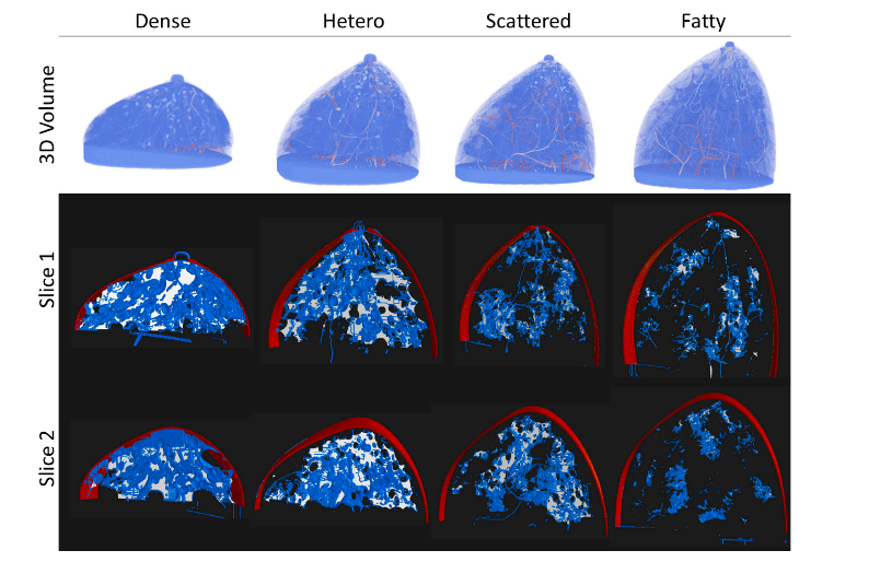
Fig. 6. Row 1: 3D renderings of VICTRE phantom breasts of four distinct profiles. Rows 2 and 3: two slices at different positions of the corresponding breast phantoms. Red: 1-voxel width breast outline; blue: extracted topological structures; white: ground truth breast tissues. Each slice’s rendering includes several additional slices around the target cross sections for detailed examination.
图 6. 第一行:VICTRE乳腺仿体的四种不同剖面的三维渲染。第二、三行:对应乳腺仿体在不同位置的两张切片。红色:1像素宽的乳腺轮廓;蓝色:提取的拓扑结构;白色:乳腺组织的真实值。每张切片的渲染还包括目标横截面周围的多个额外切片以供详细检查。

Fig. 7. Qualitative comparison of patients with and without pCR. First column: Slices of breast DCE-MRIs with tumor masked in orange (tumor masks are not used in TopoTxR). Columns 2–4: 3D renderings of topological structures from three different views. 1-D structures (loops) are rendered in blue and 2-D structures (bubbles) in red. Right: cumulative density function of topological structures’ birth times.
图 7. 对pCR(病理完全缓解)患者和未达到pCR患者的定性比较。第一列:乳腺DCE-MRI的切片,肿瘤区域用橙色遮罩(TopoTxR未使用肿瘤遮罩)。第二至第四列:从三个不同视角展示的拓扑结构三维渲染。1维结构(环)用蓝色表示,2维结构(气泡)用红色表示。右侧:拓扑结构生成时间的累积分布函数。

Fig. 8. Attention maps of CNNs when making decisions about pCR predictions. Red: voxels contributing to decisions of CNNs; blue: voxels corresponding to extracted topological structures. Columns 1, 3, 5, and 7 display 3D renderings of the attention maps, while columns 2, 4, 6, and 8 present cross-sections corresponding to the 3D renderings on their left.
图 8. CNN在进行pCR预测决策时的注意力图。红色:对CNN决策有贡献的体素;蓝色:提取的拓扑结构对应的体素。第1、3、5和7列显示注意力图的三维渲染,第2、4、6和8列显示与左侧三维渲染对应的横截面视图。
Table
表

Table 1Average percentage of each tissue type over 25 samples from each phantom breast profile.
表 1 每种组织类型在每个乳腺仿体剖面中25个样本的平均百分比。

Table 2 T1/T2 values and standard deviations for each breast tissue type
表 2 各乳腺组织类型的T1/T2值及其标准差。

Table 3Evaluation of the approximation quality of topological structures using topological precision and recall.
表 3使用拓扑精确度和召回率评估拓扑结构的近似质量。

Table 4Evaluation of the approximation quality of topological structures based on the mean distance from topological structures to breast tissues and from breast tissues to topological structures
表 4基于拓扑结构到乳腺组织的平均距离和乳腺组织到拓扑结构的平均距离对拓扑结构近似质量的评估。

Table 5Comparative analysis of our proposed method, TopoTxR, with baseline methods across four metrics – accuracy, AUC, specificity, and sensitivity – on the I-SPY1 dataset, utilizing 10-fold cross-validation. The performance of TopoTxR integrated with non-imaging clinical features (denoted as TopoTxR+Clinical) is displayed in the final row of the table.
表 5在I-SPY1数据集上使用10折交叉验证,比较我们提出的方法TopoTxR与基线方法在四项指标(准确率、AUC、特异性和敏感性)上的表现。表格最后一行显示了整合非影像学临床特征的TopoTxR(表示为TopoTxR+Clinical)的性能。

Table 6Comparative analysis of our proposed method, TopoTxR, with baseline methods across four metrics – accuracy, AUC, specificity, and sensitivity – on the proprietary Rutgers dataset, utilizing 10-fold cross-validation.
表 6在专有的Rutgers数据集上使用10折交叉验证,比较我们提出的方法TopoTxR与基线方法在四项指标(准确率、AUC、特异性和敏感性)上的表现。

Table 7TopoTxR pCR prediction results for four different densities of fibroglandular tissue in the Rutgers dataset.
表 7TopoTxR在Rutgers数据集中针对四种不同纤维腺体组织密度的pCR预测结果。

Table 8Ablation study results. All numbers are reported based on a 10-fold cross-validation on the I-SPY1 dataset.
表 8消融研究结果。所有数据均基于I-SPY1数据集的10折交叉验证得出。