Title
题目
Anatomically plausible segmentations: Explicitly preserving topology through prior deformations
解剖学上合理的分割:通过先验变形显式保持拓扑结构
01
文献速递介绍
进行环向应变或壁厚度的计算,这些测量通常用于诊断肥厚性心肌病(Brady等,2023;Corona-Villalobos等,2013),如果存在拓扑错误,这些测量的准确性可能会非常不可靠。在本研究中,我们通过连续变形具有所需拓扑特征的简化表示来划分复杂结构,以确保最终的分割具有正确的拓扑结构。
在过去十年中,耗时的手动分割任务正迅速被包括深度卷积神经网络(CNNs)在内的自动化计算工具所取代,这些工具现已成为大多数新开发的分割算法的基础(Clough等,2020;Litjens等,2017)。传统上,CNN通过对图像中的每个像素分配类别概率来执行分割任务,这一过程被称为语义分割。深度CNN对输入图像应用一系列卷积,学习局部和全局图像特征,以便基于像素的邻域进行预测。然而,由于这些网络通常使用像素级损失函数(如Dice损失或二元交叉熵(BCE))进行训练和评估,高阶结构信息往往被忽略,这可能导致解剖学上不合理的分割结果。
Abatract
摘要
Since the rise of deep learning, new medical segmentation methods have rapidly been proposed with extremelypromising results, often reporting marginal improvements on the previous state-of-the-art (SOTA) method.However, on visual inspection errors are often revealed, such as topological mistakes (e.g. holes or folds), thatare not detected using traditional evaluation metrics. Incorrect topology can often lead to errors in clinicallyrequired downstream image processing tasks. Therefore, there is a need for new methods to focus on ensuringsegmentations are topologically correct. In this work, we present TEDS-Net: a segmentation network thatpreserves anatomical topology whilst maintaining segmentation performance that is competitive with SOTAbaselines. Further, we show how current SOTA segmentation methods can introduce problematic topologicalerrors. TEDS-Net achieves anatomically plausible segmentation by using learnt topology-preserving fields todeform a prior. Traditionally, topology-preserving fields are described in the continuous domain and beginto break down when working in the discrete domain. Here, we introduce additional modifications that morestrictly enforce topology preservation. We illustrate our method on an open-source medical heart dataset,performing both single and multi-structure segmentation, and show that the generated fields contain no foldingvoxels, which corresponds to full topology preservation on individual structures whilst vastly outperforming theother baselines on overall scene topology.
自深度学习兴起以来,新的医学分割方法迅速涌现,往往取得了极具前景的成果,通常在前沿方法的基础上略有改进。然而,通过视觉检查往往会发现错误,例如拓扑错误(如孔洞或折叠),这些错误在传统的评估指标中无法检测到。拓扑结构不正确通常会导致临床上所需的下游图像处理任务出错。因此,迫切需要新的方法来确保分割结果在拓扑上是正确的。在这项工作中,我们提出了TEDS-Net,一种能够保持解剖学拓扑结构的分割网络,同时保持与前沿基线方法竞争的分割性能。此外,我们展示了当前的前沿分割方法如何引入有问题的拓扑错误。TEDS-Net通过使用学习到的拓扑保持场来变形先验,从而实现解剖学上合理的分割。传统上,拓扑保持场是在连续域中描述的,但在离散域中工作时会开始失效。为此,我们引入了额外的修改,更严格地执行拓扑保持。我们在一个开源的医学心脏数据集上展示了我们的方法,进行了单一结构和多结构的分割,并显示生成的场没有折叠体素,这意味着在单个结构上实现了完全的拓扑保持,同时在整体场景拓扑上远远超过了其他基线方法。
Method
方法
The overall aim of TEDS-Net is to automatically segment an anatomical structure of interest, whilst preserving its known topology. Toachieve this, topology-preserving fields are learnt from an input imageand used to deform a prior shape that has the desired topologicalcharacteristics, to generate a segmentation. In this section, we firstdiscuss the generation of topology-preserving fields in the discretedomain (Section 3.1), before describing how they are integrated intoTEDS-Net’s architecture (Section 3.2).
TEDS-Net的总体目标是自动分割感兴趣的解剖结构,同时保持其已知的拓扑结构。为实现这一目标,从输入图像中学习拓扑保持场,并用于变形具有所需拓扑特征的先验形状,以生成分割结果。在本节中,我们首先讨论在离散域中生成拓扑保持场(第3.1节),然后描述它们如何集成到TEDS-Net的架构中(第3.2节)。
Conclusion
结论
In this work we present TEDS-Net, a segmentation network thatdelineates structures of interest by deforming a prior shape usinglearnt topology-preserving fields. This results in anatomically plausiblesegmentations, which are crucial in many downstream medical imagingpipelines. TEDS-Net achieved 100% topology preservation across thesingle-class medical imaging tasks, whilst the Dice and HD remainedcompetitive to baseline performance.
在本研究中,我们提出了TEDS-Net,一种通过使用学习到的拓扑保持场变形先验形状来划分感兴趣结构的分割网络。此方法生成的分割结果在解剖学上合理,这在许多下游医学成像流程中至关重要。TEDS-Net在单类别医学成像任务中实现了100%的拓扑保持,同时Dice分数和Hausdorff距离(HD)也保持在与基线性能相竞争的水平。
Results
结果
On the MNIST dataset, TEDS-Net achieved an average Dice scoreof 0.90 ± 0.04 across all digits, with qualitative examples shown inFig. 7. By comparison, the U-Net achieved 0.96 ± 0.02 and VoxelMorph0.82 ± 0.06. As this segmentation task is relatively simple, it is unsurprising that the U-Net significantly (𝑝 < 0.001, as measured with apaired t-test) outperforms TEDS-Net as pixel labelling for such task ismuch simpler than deforming the prior. Moreover, this experiment waspredominantly used to assess the behaviour of TEDS-Net with differentpriors, rather than to achieve optimal Dice performance. TEDS-Netsignificantly (𝑝 < 0.001) outperformed VoxelMorph, with VoxelMorphoften found to produce bulky segmentations. It should be noted thatVoxelMorph is optimised for 3D registration not 2D segmentation asperformed in this task, however, we included this comparison to showhow other deformation methods perform with simple priors.
在MNIST数据集上,TEDS-Net在所有数字上的平均Dice分数为0.90 ± 0.04,定性示例如图7所示。相比之下,U-Net的得分为0.96 ± 0.02,而VoxelMorph的得分为0.82 ± 0.06。由于这一分割任务相对简单,U-Net显著优于TEDS-Net (𝑝 < 0.001,使用配对t检验测量),这一结果并不令人惊讶,因为对于这种任务,像素标记比变形先验要简单得多。此外,这个实验主要用于评估TEDS-Net在不同先验条件下的表现,而不是为了获得最佳的Dice分数。TEDS-Net显著优于VoxelMorph (𝑝 < 0.001),因为VoxelMorph常常产生过于庞大的分割。需要注意的是,VoxelMorph优化用于3D配准而不是本任务中的2D分割,然而我们仍然包含了这个比较,以展示其他变形方法在简单先验下的表现。
Figure
图

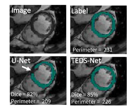
Fig. 1. Example of topological errors found when segmenting medical images using traditional CNNs, i.e. a U-Net style architecture, trained with a pixel-wise loss function. Here we show myocardium segmentations from 2D MRI short axis slices. The gap found within the U-Net example (indicated with the white arrow) changes the topology and interferes with automated measures of perimeter.
图1. 使用传统卷积神经网络(CNNs)进行医学图像分割时出现的拓扑错误示例,即使用像素级损失函数训练的U-Net风格架构。在这里,我们展示了2D MRI短轴切片中的心肌分割。U-Net示例中发现的间隙(用白色箭头指示)改变了拓扑结构,并干扰了自动化的周长测量。

Fig. 2. Panels A-D. Panel A shows a simple 1D transform Φ1 and the composition (one application of ‘‘scaling and squaring’’) obtained through both discrete (solid line) and continuous (dotted line) methods. The grey arrows illustrate the two steps used to calculate Φ2 in the discrete case at 𝑥 = 1, Φ2 (1) = Φ1 (Φ1 (1)) = Φ1 (1.5) = 2. Panel B shows a series of composition layers and Panel C and D the same transforms after using more finely sampled points, with and without Gaussian smoothing.
图2. 面板A-D。面板A展示了一个简单的一维变换 Φ1 以及通过离散(实线)和连续(虚线)方法获得的组合(“缩放和平方法”一次应用)的结果。灰色箭头说明了在离散情况下在𝑥 = 1处计算 Φ2 的两个步骤,Φ2 (1) = Φ1 (Φ1 (1)) = Φ1 (1.5) = 2。面板B展示了一系列组合层,而面板C和D则展示了使用更细采样点后的相同变换,分别在有和没有高斯平滑的情况下。

Fig. 3. Schematic diagram of TEDS-Net, illustrated for multi-structure segmentation with 𝑐 channels. Two deformation fields are learnt through a series of convolutions appliedto an input image, before being encouraged to be topology-preserving through our topology-preserving layers (shown in the green box).
图3. TEDS-Net的示意图,展示了用于多结构分割的 𝑐 个通道。通过对输入图像应用一系列卷积学习到两个变形场,然后通过我们的拓扑保持层(绿色框中所示)进行调整,以保持拓扑结构。

Fig. 4. Visualisation of the scaling and squaring approach, shown in one direction. Initially, the fields have negligible displacements, but after composing it by itself ℎ = 8 times(Φ2 ℎ = Φ256) the displacements are amplified.
图4. 缩放和平方法的可视化,显示了一个方向的变化。最初,这些场的位移可以忽略不计,但经过自我组合 ℎ = 8 次(Φ2 ℎ = Φ256)后,位移被放大。

Fig. 5. An overview of the priors used for each experiment. Panel A shows the priorsused for each of the MNIST digits and Panel B for myocardium experiments. PanelC shows the set of priors used for multi-structure cardiac segmentation and theircorresponding label. Further, this figure shows the density of the right ventricle (rv)label in the train, validation and test datasets before augmentation.
图5. 每个实验中使用的先验概览。面板A展示了用于每个MNIST数字的先验,面板B展示了用于心肌实验的先验。面板C展示了用于多结构心脏分割的先验集合及其对应的标签。此外,该图还显示了在增强之前训练、验证和测试数据集中右心室(rv)标签的密度。

Fig. 6. Segmentation of the heart shown in 3D and at two 2D cross sections in theshort-axis. Depending on the location of the short-axis slice, the topology of eachstructure can vary. For TEDS-Net the topology must be known and therefore, onlyslices with the same topology as shown in Slice A were used across all experiments.
图6. 心脏的分割结果在3D和两个2D短轴截面上的展示。根据短轴切片的位置,每个结构的拓扑结构可能会有所不同。对于TEDS-Net来说,拓扑结构必须是已知的,因此在所有实验中只使用了与切片A所示相同拓扑结构的切片。

Fig. 7. MNIST digit segmentations using TEDS-Net. The blue lines show the labels (𝐘), whilst the red show TEDS-Net predictions ( ̂𝐘).
图7. 使用TEDS-Net进行MNIST数字分割的结果。蓝色线条表示标签(𝐘),红色线条表示TEDS-Net的预测结果(̂𝐘)。

Fig. 8. Examples of where TEDS-Net has not preserved the topology of the prior, theseexamples were randomly chosen.
图8. TEDS-Net未能保持先验拓扑结构的示例,这些示例是随机选择的。

Fig. 9. The effect that 𝜎 within the Gaussian smoothing kernel played on segmentationperformance, shown with the average Dice score and Hausdorff distance. The bottomrow shows 𝜎’s role in preventing folding voxels, where folding voxels are given by% 𝐽*Φ ≤ 0 in the bulk (left) and fine-tuning branch (right).
图9. 高斯平滑核内的 𝜎 对分割性能的影响,显示了平均Dice分数和Hausdorff距离。底部显示了 𝜎 在防止折叠体素中的作用,其中折叠体素由% 𝐽*Φ ≤ 0 在主体(左侧)和微调分支(右侧)中给出。

Fig. 10. Qualitative performance of myocardium segmentation for TEDS-Net and across the baselines for four different patients (a-d). The ground truth labels (𝐘) and predictedsegmentations ( 𝐘) are shown in green and red, respectively.
图10. TEDS-Net及其基线方法在心肌分割上的定性表现,针对四位不同患者(a-d)的结果。绿色表示真实标签(𝐘),红色表示预测分割结果(𝐘)。

Fig. 11. The relative error of automated myocardium perimeter measurements acrossthe five methods. This shows the ratio between the absolute error and the knownperimeter value, expressed as a percentage of the known perimeter value. The averagemyocardium perimeter was found to be approximately 250 voxels. The myocardiumperimeter from each prediction was compared to the perimeter of the labels. Themean for each network is shown in white, and predictions with incorrect topologyare highlighted.
图11. 不同方法在自动化心肌周长测量中的相对误差。图中显示了绝对误差与已知周长值之间的比率,以已知周长值的百分比表示。心肌的平均周长大约为250个体素。每个预测的心肌周长与标签的周长进行了比较。每个网络的平均值以白色显示,拓扑结构不正确的预测结果被突出显示。

Fig. 12. The segmentation performance when 𝐏𝑟𝑣 was at each position defined in Fig. 5C, performed on the validation set.
图12. 当 𝐏𝑟𝑣 处于图5C中定义的每个位置时的分割性能,基于验证集进行的测试结果。

Fig. 13. Qualitative examples to show TEDS-Net over-segmenting in regions of thinboundaries. The ground truth is shown in green, TEDS-Net predictions in red andexamples at different 𝜎’s (𝜎 = 0 is no smoothing). Arrows have been added in thesame position to highlight the segmentations mistakes at the thinnest regions of thelabel.
图13. TEDS-Net在薄边界区域过度分割的定性示例。图中绿色表示真实标签,红色表示TEDS-Net的预测结果,显示了在不同 𝜎 值下的示例(𝜎 = 0 表示没有平滑)。在相同位置添加了箭头,以突出标签最薄区域的分割错误。
Table
表

Table 1Summary of each network’s performance at segmenting the myocardium from the ACDC dataset. The bestresult (but not significant) is highlighted in bold. Hausdorff distance (HD) is measured in mm. The trainingtime per epoch and number of parameters used for each method is also reported.
表1 各网络在ACDC数据集中分割心肌的性能总结。最佳结果(但不显著)以粗体显示。Hausdorff距离(HD)以毫米为单位测量。同时报告了每种方法的每个训练周期的时间和所使用的参数数量。

Table 2Segmentation performance and topology preservation rates between TEDS-Net and the baselines, on eachstructure: the right ventricle (rv), the myocardium (myo) and left ventricle (lv) as well as the overall scene.The best performance for each measure is highlighted in bold.
表2 TEDS-Net与基线方法在每个结构上的分割性能和拓扑保持率的比较:右心室(rv)、心肌(myo)和左心室(lv),以及整体场景的表现。每项指标的最佳性能以粗体显示。